Introduction
Chondroid lesions, also known as cartilaginous tumours, are a diverse group of neoplasms or growths that primarily affect cartilage. These lesions can occur in various locations throughout the body, most commonly in the bones and soft tissues, and can range from benign (harmless) to malignant (cancerous) forms. Understanding the symptoms, diagnostic procedures, and treatment options for chondroid lesions is essential for effective management and improved patient outcomes. This article will delve into the various aspects of chondroid lesions, particularly in the context of the Manchester Lumps Clinic, a leading facility for diagnosing and treating such tumours.
Symptoms of Chondroid Lesions
Common Symptoms
Chondroid lesions are commonly asymptomatic but can present with a variety of symptoms, often depending on their location and size. Often the symptoms are due to other reasons near the joint or bone.
Common symptoms include localized pain, swelling, and tenderness in the affected area. Patients may also experience limited range of motion if the lesion is located near a joint. In cases where the lesion is larger, it may cause noticeable deformity in the affected limb or area. Additionally, patients may report discomfort during physical activities, leading to decreased functionality and quality of life.
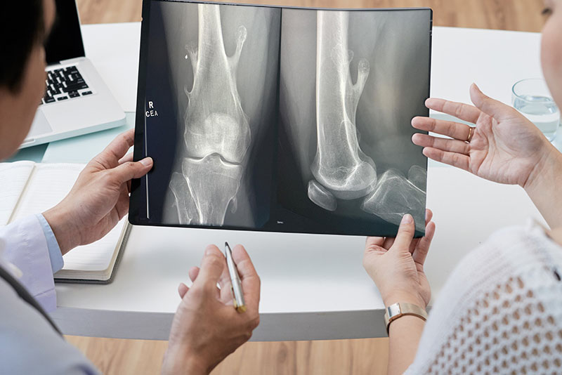
However, it is more common for such chondroid lesions within bone to be picked up incidentally on investigations on the bone such as X-rays or MR scans for other reasons. The common areas where they are seen are in the fingers (e.g. Enchondroma), the thigh bone such as a femur, and also in the pelvis.
In some instances, especially with malignant (harmful) chondroid lesions, systemic symptoms such as fever, weight loss, and fatigue may occur. These symptoms can be indicative of an aggressive tumour and warrant immediate medical evaluation. Understanding these signs is crucial for early identification and timely intervention, ultimately improving prognosis and treatment success.
Potential Complications
If left untreated, chondroid lesions can lead to several complications, particularly if they are malignant. They can be locally destructive if growing in the bone and one potential complication is the risk of metastasis, where cancerous cells spread to other parts of the body, leading to further health issues. Additionally, benign lesions can still cause significant complications by compressing surrounding structures, resulting in nerve damage, vascular compromise, or joint dysfunction. Early diagnosis and treatment are essential to minimize these risks and address the lesion effectively.
It is imperative for healthcare providers to monitor patients with chondroid lesions closely and intervene promptly to prevent these complications from arising.
Diagnostic Procedures
Initial Assessment
The journey towards diagnosing chondroid lesions typically begins with a thorough medical history and physical examination. Healthcare providers will enquire about the patient’s symptoms, duration, and any previous medical conditions that might be relevant. During the physical examination, the provider may assess the affected area for swelling, tenderness, and range of motion, which can provide valuable clues regarding the nature of the lesion.
After the initial assessment, if a chondroid lesion is suspected, the provider will likely refer the patient for further imaging studies to gain a more comprehensive understanding of the lesion’s characteristics and extent.
Imaging Techniques
Imaging techniques play a crucial role in the diagnosis of chondroid lesions. X-rays are typically the first imaging modality used, offering a basic view of the bone structure and any visible lesions. However, for more detailed assessment, advanced imaging techniques such as Magnetic Resonance Imaging (MRI) and Computed Tomography (CT) scans are often employed. MRI is particularly useful for evaluating soft tissue components and determining the extent of the lesion.
CT scans provide detailed images of the bone architecture and can help identify any cortical involvement or associated bone marrow oedema. The combination of these imaging techniques allows for a more accurate characterisation of the lesion, which is essential for determining the appropriate management strategy.
Biopsy Procedures
In many cases, imaging alone is not sufficient to determine the nature of a chondroid lesion, necessitating a biopsy for definitive diagnosis. Biopsy is performed usually by core needle biopsy to obtain tissue for an accurate histopathological diagnosis.
Excisional biopsy, although more invasive, allows for complete removal of the lesion along with surrounding tissue, providing both diagnostic and therapeutic benefits. The tissue obtained during biopsy is then sent to a pathologist for histological examination, which is critical for determining the lesion’s nature – benign, malignant, or otherwise.
Treatment Options Available
Conservative Management
The treatment approach for chondroid lesions largely depends on the lesion’s type, location, symptoms, and whether it is benign or malignant. For benign chondroid lesions that are asymptomatic and not causing significant issues, conservative management may be appropriate. This can include regular monitoring through follow-up imaging and clinical evaluations. In some cases, the use of pain relievers or physical therapy may help alleviate discomfort associated with the lesion.
However, if conservative measures fail or if the lesion begins to cause complications, more aggressive treatment options may be necessary. It is essential for healthcare providers to maintain open communication with their patients regarding the best management strategies tailored to individual circumstances.
Interventional Procedures
For symptomatic or malignant chondroid lesions, interventional procedures are often warranted. Surgical excision is the primary treatment modality for both benign and malignant lesions. The goal of surgical intervention is to remove the tumor completely while preserving surrounding healthy tissue whenever possible. In cases of malignant chondroid lesions, wider excisional margins may be necessary to ensure that no cancerous cells are left behind.
In addition to surgery, adjuvant therapies such as radiation therapy or chemotherapy may be employed for malignant lesions, particularly in cases where there is a risk of metastasis. These treatments can be pivotal in managing aggressive tumors and improving overall survival rates. The multidisciplinary approach to treating chondroid lesions ensures that patients receive comprehensive care tailored to their unique needs.
Rehabilitation and Follow-Up Care
Post-surgical rehabilitation is a critical component of management for patients with chondroid lesions. Depending on the location and extent of the surgery, rehabilitation may involve physical therapy to restore strength and mobility to the affected area. Regular follow-up appointments are essential to monitor for any recurrence of the lesion or development of new symptoms. Imaging studies may also be repeated to assess the success of treatment and ensure that the lesion has been adequately addressed.
Patients must be educated about the signs of complications, such as persistent pain or swelling, and encouraged to seek medical attention if these symptoms arise. Ongoing research into the management of chondroid lesions continues to evolve, providing new insights and treatment options for affected individuals.
Conclusion
In summary, chondroid lesions present a complex challenge for both patients and healthcare providers. Understanding the symptoms, diagnostic procedures, and available treatment options is crucial for effective management. With advancements in imaging technology and treatment modalities, the prognosis for individuals with chondroid lesions has greatly improved. Early detection and intervention remain key to optimizing outcomes and preventing complications.
As research in this field continues to progress, it is vital for patients experiencing symptoms related to chondroid lesions to seek evaluation and care from specialized medical professionals, such as those at the Manchester Lumps Clinic. This approach ensures that they receive appropriate and timely treatment, ultimately enhancing their quality of life.